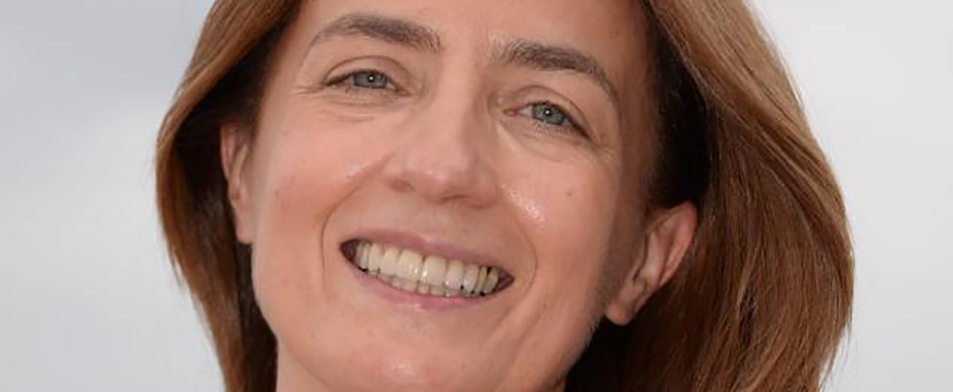
Prof. Antonia Carla Testa - Scientific Director of the Center for Ultrasound in Gynecological Oncology “Class Ultrasound” of the Agostino Gemelli University Hospital

Where did the idea that an ultrasound scan could detect Covid 19 Pneumonia come from?
I have been carrying out research into the use of ultrasound examinations for the assessment of gynaecological tumours for many years and a few months ago I invited a colleague, the paediatrician Dr Danilo Bonsenso, to explain to my residents how he uses ultrasound scans of the lungs in the paediatric field. When the pandemic was announced, one young doctor asked me if I had ever heard of ultrasound scans of the lungs and I immediately thought of Dr Bonsenso. I realized that lung ultrasound examination was an accurate imaging method to diagnose Covid-19 pneumonia.
My first thoughts went to pregnant women, for whom it is not easy to undergo one or more Computed Tomography examinations. Then I thought of the many gynaecologists working around the world and already experts in the use of ultrasound scanning: perhaps it would have been possible to teach a person already experienced in the technique to evaluate another organ. In just 24 hours we prepared a four-minute video tutorial which was published in the Internation Journal, Ultrasound in obstetrics and gynaecology.
A task force was immediately set up, which included Dr Bonsenso and two doctors Smargiassi and Inchignolo who, in cooperation with the University of Trento, had developed an Artificial Intelligence software and a platform capable of receiving the clips of ultrasound scans from various parts of the lung with the possibility to automatically detect any abnormalities.
We developed a fast teaching program to verify whether this technique could be easily learned and it was found to be possible and effective. Then someone mentioned Africa. Thinking of the African nations who, fortunately, were not yet affected by this storm but were preparing to face an emergency, we realized that there are many places such as hospitals or missionary centres which are unlikely to have a CT scanner, but might well have an ultrasound scanner. Speaking to the task force, I suggested that we shared the idea with our African colleagues. CESI played an important role in making it possible for us to organize the project. We contacted English-speaking African nations in various ways and within ten days we had set up two webinar appointments. Our main intention was not to contact our colleagues in Africa with the attitude of those who want to teach, but to set up a cooperative project involving genuine sharing.
How long did it take to set up the project?
It took less than two months and the teamwork was extremely important, in particular the work with my closest colleague, Dr Moro. The work was relentless but thrilling. The best thing is that we have involved young people in the project and this group of residents who welcomed my proposal played a key role in the two days of the project. Once we realized that twelve nations would have been involved, we thought there might have been problems with the Internet connection, so in advance of the meeting we set up WhatsApp chats for each resident with a specific nation so that they could have been in personal contact. We wanted the explanatory part to be as long as needed, practical and interactive, with a self-evaluation quiz. Then we invited our African colleagues – doctors, nurses, midwives and sonographers – to provide feedback in a session, called Voices from Africa. At first the connection was problematic, but once the first colleague appeared on the screen and told us how they were coping with the situation, there was a welcoming round of applause and that was our reward for the effort of the past weeks.
How rewarding was this experience for you, as doctors?
As everyone struck by of this crisis initially we felt quite impotent and this opportunity to act certainly energised us and allowed us to say, ‘we don’t know if it will work, but let’s try’. Working directly and interactively with the young residents meant that we felt their enthusiasm and we were able to engage them in the project and the opportunity to help other colleagues and ourselves to explore areas outside our specific microcosms. Knowing that we were helping a colleague on the other side of the world made our work and our skills all the more valuable. We often use the expression “if we all pull together…” and we were able to involve others in this adventure, giving them tasks to do, stimulating their creativity, and when a colleague in the intensive-care ward needed an ultrasound scan of a lung, we could make ourselves useful; we could use our specific skills to soothe a pregnant woman about the health of her lungs. Finally, the link with Africa was really special for me.
You gave your know-how, what did you receive in return?
When you give, you always receive: we are a multidisciplinary group and we were provided with a tool that will be very useful for our cancer patients. We made a number of contacts with staff in Africa, which will certainly continue. We felt that we were involved in our mission: we talk a lot about the third mission of Università Cattolica and so we are very much aware that clinical medicine comes first, but it is also our duty to carry out research, exactly because we are a university. Moreover, it is equally important to educate our young people and to offer the knowledge and results generated in our laboratories not only to the outside community here in Italy, but also to those who live overseas.
What additional skills are needed when the relationship is mediated by technology?
We were all a little hampered by the lack of physical presence and closeness at the beginning. However, I saw that these instruments are amazing when they are used correctly. If a colleague has difficulty with the Internet connection, you wait, you watch, you listen. Although you are not present to take their hand and guide them in using the probe, you can demonstrate and you can get them to repeat the operation. I found that - like so many things - if technologies are used correctly, they have enormous potential. The work we did with Africa was important. I had known little about that world and this reciprocal learning situation was marvellous: getting ready to understand their situation, especially listening to their experiences, and giving them back our suggestions with a sense of humility and collaboration.

